Carneige stage 2 - 1st Trimester
Cleavage - 1st Cell Division
•••••
SIZE: 0.1 - 0.2 mm
TIME PERIOD: 1.5 - 3 days post-ovulation
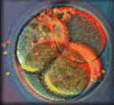
The zygote now begins to cleave, with each division occurring into two cells called blastomeres. The zygote's first cell division begins a series of divisions, with each division occurring approximately every twenty hours. Each blastomere within the zona pellucida becomes smaller and smaller with each subsequent division.
When cell division generates about sixteen cells, the zygote becomes a morula (mulberry shaped). It leaves the fallopian tube and enters the uterine cavity three to four days after fertilization.
Carneige stage 3 - 1st Trimester
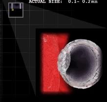
Early Blastocyst
•••••
SIZE: 0.1 - 0.2 mm
TIME PERIOD: 4 days post - ovulation

About four days after fertilization, the morula enters the uterine cavity. Cell division continues, and a cavity known as a blastocele forms in the center of the morula. Cells flatten and compact on the inside of the cavity while the zona pellucida remains the same size. With the appearance of the cavity in the center, the entire structure is now called a blastocyst.
The presence of the blastocyst indicates that two cell types are forming: the embryoblast (inner cell mass on the inside of the blastocele), and the trophoblast (the cells on the outside of the blastocele).
Carneige stage 4- 1st Trimester
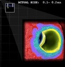
Implantation begins - hCG Levels Rise
•••••
SIZE: 0.1 - 0.2 mm
TIME PERIOD: 5 - 6 days post-ovulation
The pressure of the blastocele expanding in the middle of the blastocyst against the rigid wall of the zona pellucida, creates "hatching" of the blastocyst from the zona around the sixth day after fertilization.
As the blastocyst enters the uterus free from the zona, the outer layer of trophoblast cells secrete an enzyme to erode the epithelial lining of the uterus and allow the blastocyst to implant.
Human chorionic gonadotropin (hCG) is also secreted by the trophoblast cells stimulating the corpus luteum to continue progesterone production. Progesterone maintains the blood rich lining of the uterus. Endometrial glands in the uterus had already begun to enlarge in response to the progesterone stimulated to release by the corpus luteum ("yellow body") which once surrounded the egg as it grew in the ovary almost six days prior.
The uterus is therefore swollen with new blood capillaries and the circulation between mother and blastocyst begins, a process needed for the continuation of pregnancy.
Carneige stage 5 - 1st Trimester
Implantation Complete
•••••
SIZE: 0.1 - 0.2 mm
TIME PERIOD: 7 - 12 days post-ovulation
Trophoblast cells continue to engulf and destroy cells of the uterine lining creating blood pools and stimulating new capillaries to grow - beginning the growth of the placenta.
The blastocyst inner cell mass differentiates into two layers:
EPIBLAST The top layer of cells (dark blue) which will become the embryo and amniotic cavity.
HYPOBLAST Lower layer of cells (yellow) which become the yolk sac.
Carneige stage 6 - 1st Trimester
Primitive Streak
•••••
SIZE: 0.2 mm
TIME PERIOD: 13 days post-ovulation
 Placenta Formation
Chorionic villi "fingers" form in the "placenta" anchoring the embryo to the uterus. Blood vessels begin appearing first in the "placenta" surrounding the embryo. The yolk sac begins producing hematopoietic or non-nucleated blood cells.
Stalk Formation
By the end of stage 6a, the embryo is attached to the developing placenta by a stalk later to become part of the umbilical cord.
Gastrulation
In Carnegie Stage 1, the exit of the first polar body through the zona pellucida determined the gastrulation axis.
Stage 6b begins as a narrow line of cells appear on the surface of the formerly 2 layered embryonic disc. This "primitive streak" marks bilateral symetry in the embryo and indicates gastrulation has begun - the migration of cells from the outer edges of the disc into the primitive streak and down creating a new third layer. The embryo now has endoderm, mesoderm and ectoderm layers.
ECTODERM
Top cell layer of the embryonic disc. Will later form: skin, hair, lenses of the eyes, lining of the internal and external ear, nose, sinues, mouth, anus, tooth enamel, pituitary and mammary glands, and all parts of the nervous system.
MEDODERM
Middle cell layer of the embryonic disc and precursor to the muscles, bones, lymphatic tissue, spleen, blood cells, heart, lungs, reproductive and excretory systems.
ENDODERM
Inner cell layer of the embryonic disc from which will form the lining of the lungs, the tongue, tonsils, urethra and associated glands, bladder and digestive tract.
Carneige stage 7 - 1st Trimester
Neurulation
•••••
SIZE: 0.4 mm
TIME PERIOD: 16 days post-ovulation
 Neurulation
In Stage 6, gastrulation began with the appearance of the primitive streak. In Stage 7, gastrulation continues with the formation of a new cell layer - the ectoderm - changing the two-layered disc into a three-layered disc.
Nerual crest cells originate at the top of the neural tube and migrate extensively, differentiating into many cell types such as neurons, glial cells, pigmented cells of the epidermis, epinephrine producing cells of the adrenal gland, and various skeletal and connective tissues of the head. It appears that the fate of a migrating cell is largely determined by its final destination.
RhoB and Slug protein are the proteins now present in Stage 7 which promote migration. The loss of N-cadherin, a protein required for left-right asymmetry, also helps to initiate the migration of neural crests cells.
Gastrulation
Gastrulation in full force. Cells are migrating across the surface of the 2 layered embryonic disc and into the newly formed Primitive Streak. After migrating through the Primitive Streak, these new cells collect into a new 3rd layer - the endoderm.
Carneige stage 8 - 1st Trimester
Neural groove
•••••
SIZE: 1.0 - 1.5 mm
TIME PERIOD: 17-19 days post-ovulation
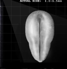
The embryonic area is now shaped like a pear, and the head region is broader than the tail end.
The ectoderm has thickened to form the neural plate. The edges of this plate rise and form a concave area known as the neural groove. This groove is the precursor of the embryo's nervous system and it is one of the first organs to develop.
By stage 8, the blood cells are already developed and begin to form channels along side of the epithelial cells forming at the same time.
Carneige stage 9 - 1st Trimester
Appearance of Somites
•••••
SIZE: 1.5 - 2.5 mm
TIME PERIOD: 19 - 21 days post-ovulation
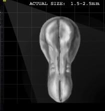
Looking at the embryo from the top, the head end is wider than the tail end, with a slightly narrowed middle.
Somites, which are composed of mesoderm, appear on either side of the neural groove. The first pair of somites appear at the tail and progress to the middle. One to three pairs of somites are present by Stage 9.
Every ridge, bump and recess now indicates cellular differentiation.
A head fold rises on either side of the primitive streak. The primitive streak now runs between one-fourth to one-third of the length of the embryo.
Secondary blood vessels now appear in the chorion/placenta. Hematopoietic cells appear on the yolk sac simultaneously with endothelial cells which will go on to form blood vessels for the newly emerging blood cells.
Endocardial (muscle) cells begin to fuse and form into the early embryo's two heart tubes.
Carneige stage 10- 1st Trimester
Neural Folds/Heart Folds Begin to Fuse
•••••
SIZE: 1.5 - 3.0 mm
TIME PERIOD: 21- 23 days post-ovulation
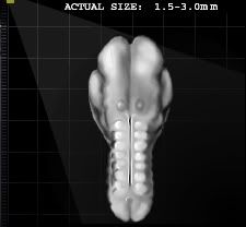
Rapid cellular growth and change elongates the embryo and expands the yolk sac.
On each side of the neural tube, between four and twelve pairs of somites can exist by the end of Stage 10. The cells which become the eyes appear as thickened circles just off of the neural folds. The newly differentiated cells of the ears are also present.
Neural folds are rising and fusing at several points along the length of the neural tube concomitant with the budding somites which appear to "zipper" the neural tube closed. Neural crest cells will eventually contribute to the skull and face of the embryo.
The two endocardial tubes formed in Stage 9 now fuse in Stage 10. Together they form one single tube generated from the cells of the "roof" of the nueral tube. The heart tube takes on an S-shape establishing the asymetry of the heart. As the S-shape forms, cardiac muscle contraction begins.
Carneige stage 11- 1st Trimester
Two Pharyngeal Arches Appear
•••••
SIZE: 2.5 - 3.0 mm
TIME PERIOD: 23- 25 days post-ovulation
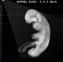
Thirteen to twenty pairs of somites are present and the embryo is shaped in a modified S curve. The embryo has a bulb-like tail and a connecting stalk to the developing placenta.
A primitive S-shaped tubal heart is beating and peristalsis, the rhythmic muscle contractions propelling fluids throughout the body, begins. However, this is not true circulation because blood vesel development is still incomplete.
At this stage, the neural tube determines the form of the embryo. Although the primary blood vessels along the central nervous system are now connecting in Stage 11, the central nervous system appears to be the most developed system. If twenty somites are present in the embryo, the forebrain is completely closed.
 and it's
  news today
 .
.
 to all.