Uterine problems
Many women who have uterine problems may have no problems getting pregnant, but they may have difficulty in keeping the pregnancy as they tend to miscarry. Most women have no symptoms, some women will have scanty periods, and if the woman has no uterus she will have no periods, (but remember there are many causes of absent periods). Some patients may also complain of heavy periods (due to fibroids).
Incidence
10-15%
Causes
Fibroids
Fibroids are benign round growths of the muscle of the womb; they are very common especially in women of reproductive age. Approximately one fifth to one third of all women have fibroids. Fibroids are more common amongst some racial groups such as Africans and afro Caribbean women. They can run in families. Fibroids can vary in size from a few millimetres to that of a full term pregnancy. Fibroids also vary in location, either inside the cavity wall itself (intramural), bulging from outside wall of the womb (subserous) or bulging inside the cavity of the womb (submucousal). Fibroid can be found hanging on a stalk either outside or inside the womb (pedunculated). Cervical fibroids are fibroid in the cervix (neck of the womb). What causes fibroids is unknown. The growth of fibroid depends on the amount of hormones oestrogen and progesterone in the body and subsequently increases in size with advanced age. However they shrink after the menopause. Fibroids may also grow during pregnancy due to excess production of the oestrogen during pregnancy.
Some women with fibroids experience no symptoms at all, others may experience symptoms, such as heavy periods, heavy painful periods causing anaemia, and pressure symptoms such as feeling bloated and swollen abdomen, need to pass urine more frequently, abdominal distension, discomfort during sex, back pain and constipation, these symptoms depend on the size, number and location of fibroids. Most women who have fibroids are fertile and have no problems with keeping the pregnancy. However, if the fibroid significantly distorts the cavity of the womb, it may interfere with embryo implantation. Also fibroids can displace or block the Fallopian tubes. Most fibroids are diagnosed by ultrasound scan either through the abdomen (transabdominal scan) or through the vagina (transvaginal scan), sometimes MRI imaging may be required to add more information about the location of fiboids. Fibroids can also be diagnosed at operations such as laparoscopy and hysteroscopy. They can also be picked up by HSG.
Laparoscopy showing multiple fibroid uterus

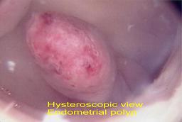
Uterine Polyps (Endometrial polyp)
Uterine polyps are small benign growths of the lining of the womb (endometrium) dangling in the cavity of the womb. Endometrium polyp are very common and can be single or multiple and can vary in size from very small with minimal distortion of the womb cavity to large one filling the whole uterine cavity measuring several cenitmetres in length. Some women with endomtrial polyp experience no symptoms at all, othere may experience irregular and sometimes heavy menstrual bleeding. Endometrial polyp can interfere with conception by acting like a coil. There is evidence that removal of large polyp prior to infertility treatment and IVF may prove the clinical pregnancy rate. In general, the significance of polyp in relation to infertility depend upon their type, size and location.
Hysteroscopy showing endomettrial polyp
Ultrasound sca showing endomettrial polyp

Ultrasound scan showing good endometrium development

Intrauterine adhesions
Intrauterine adhesions, known as Ashermans syndrome, is scar tissue inside the uterine cavity connecting parts of it. Ashermans syndrome is often caused by inflammation or damage to the lining of the womb. Asherman syndrome can cause absent periods, interfere with conception, and can increase the risk of miscarriage.
Endometrial lining
Both too thin and too thick an endometrium could reduce the chance of conception. In general 8-12 mm endometrial thickness is good when measured by ultrasound scan around day 10-12 of the cycle.There is an association between open cavity and poor outcome following IVF treatment.
Ultrasound scan showing open uterine cavity

Congenital Problems
Congenital problems (problems from birth) e.g. absent
uterus, the uterus has not developed sufficiently in size (hypoplastic
uterus). Mayer Rokitansky Kuster Hauser Syndrome where the uterus and vagina are absent or not sufficiently developed in a woman with normal chromosome 46XX. The patient usually present with absent periods despite normal development of secondary sex characterics, and or with failure to attempt vaginal intercourse. Recurrent abdominal pain is not uncommon.
Abnormally shaped uterus such as T shaped uterus, double uterus, birconuate (two-horned uterus), unicornuate (a half uterus) and uterine septum (this is the most common congenital abnormalities and is associated with the highest incidence of reproductive failure such as recurrent miscarriage and increased preterm birth).
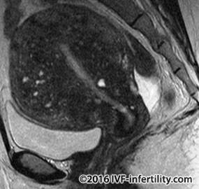
Adenomyosis
The term adenomyosis refer to a relatively common condition in which cells like the one that line the inside of the womb (endometrium) are present in the muscles of the womb. The abnormally located endometrial tissue tends to bleed with menses creating small fluid filled collections inside the womb muscles. The womb may become larger, globular and tender. Adenomyosis tends to occur in women who are in their 30s and 40s particularly if they have had children. Adenomyosis is benign condition and does not cause cancer. The cause of adenomyosis remains unknown. Most women with adenomyosis do not experience any symptoms, while others may suffer heavy painful periods and or pelvic pain during intercourse. Many women with adenomyosis will have endometriosis as well. The diagnosis is usually suspected by vaginal ultrasound which demonstrate globular large uterus, poor definition of the endometrial-myometrial interface (junction zone) and the presence of endometrial cysts. MRI can be helpful when ultrasound does not give definite results. Most commonly adenomyosis is mistaken for another common condition, uterine fibroids. The effect of adenomyosis on fertility is not clear but it may lower fertility. Puente Agueda and colleagues from Spain (2014) reported high prevalence of adenomyosis in recurrent miscarriages and previous IVF failure.
MRI showing adenomyosis in the womb
A forgotten coil
How common is the problem of forgotten coil (IUCD)? There is no published data ascertaining the frequency of forgotten IUCD. However, the author believes that the problem is probably underestimated or under reported. Although rare, it is important to consider the possibility of a forgotten IUCD as a cause of unexplained infertility (Marcus et al. Fertility Sterility, 2011).
Ultrasound showing forgotten coil inside the womb

Ultrasound showing Chinese forgotten coil inside the womb
