Ultrasound scan
The use of high frequency ultrasound waves to examine the cervix, uterus, ovaries and Fallopian tubes. The reflection of these waves can be used to produce an image on a screen similar to an X-ray, but with the added advantage that the ultrasound scan is quite harmless. Ultrasound examination is routinely performed as an outpatient procedure and takes about 10 minutes.
It is used as a diagnostic procedure to assess pelvic organs and detect abnormalities in the womb e.g. a forgotten coil (used for contraception), polyp, malformation, abnormalities in the Fallopian tubes e.g. hydrosalpinx (water in the tubes) and abnormalities in the ovaries e.g. polycystic ovaries etc.
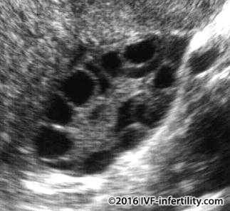
Ultrasound scan showing Polycystic ovary
Ultrasound scan showing ovarian cyst
Ultrasound scan showing hydrosalpinx
Ultrasound scan showing large polyp
Ultrasound scan showing large endometrioma.
It is also used to monitor treatments such as insemination and IVF by assessing the development of the follicles and measuring the thickness of the endometrium.
Ultrasound scan showing normal endometrium.
Ultrasound scan is the method of choice to diagnose pregnancy and multiple pregnancies. The pelvic organs can be examined either by passing the probe over the abdomen (abdominal scan) or by gently inserting the probe into the vagina (vaginal scan). For best results, the abdominal scan requires a full bladder, which pushes the bowel away from the ovaries, and at the same time acts as a good transmitter for the ultrasound waves. A vaginal scan gives a more accurate picture of the pelvic organs than an abdominal scan because of the shorter distance for the sound waves to travel (between the probe and the pelvic organs). A full bladder is not required for a vaginal scan, hence making the patient more comfortable.
Ultrasound of normal intrauterine pregnancy